Selenium, Soil Depletion, and Your Child's Developing Brain: What Every Parent Should Know
Why a mineral vanishing from farmland soil could quietly undermine thyroid-driven brain development—and what you can actually do about it.
The thyroid gland is not simply a metabolism regulator. During pregnancy and early childhood, it is essentially a brain-building organ—and it cannot do that job without selenium. Most parents have heard about iodine and thyroid health. Far fewer know that selenium is equally essential, that it is disappearing from agricultural soils across large parts of the world, and that low maternal selenium during pregnancy has been linked in prospective research to higher rates of ADHD and autism traits in children.
This is not a fringe concern. It is a mechanistic story with real human data behind it.
What the Thyroid Actually Does for a Developing Brain
Thyroid hormones—primarily thyroxine (T4) and its active form triiodothyronine (T3)—orchestrate an enormous range of neurodevelopmental processes. They direct neuronal migration, axon formation, and, critically, myelination: the wrapping of nerve fibers in the fatty insulating sheath that determines how fast and reliably signals travel (Calcaterra et al., Nutrients, 2025). When the hypothalamic-pituitary-thyroid axis is still immature in early infancy, disruptions here can produce complications that echo across years of development.
Iodine deficiency gets most of the attention in this story—and rightly so. Even mild iodine insufficiency in pregnant women and neonates impairs neurointellectual development in their children (Delange, Postgraduate Medical Journal, 2001). But iodine is only part of the picture. T4, the form the thyroid secretes, is largely inactive. It must be converted to T3 inside target tissues—including the brain—by enzymes called iodothyronine deiodinases. Those enzymes are selenoproteins. Without selenium, the conversion cannot happen properly (Köhrle & Gärtner, Best Practice & Research Clinical Endocrinology & Metabolism, 2009).
Selenium Is the Key That Unlocks Thyroid Hormone in the Brain
There are three deiodinase enzymes (DIO1, DIO2, DIO3), each embedded with the rare amino acid selenocysteine at its active site. DIO2 in particular generates the active T3 that neurons need locally—inside glial cells in the brain—independent of circulating T3 levels (Gereben et al., Cellular and Molecular Life Sciences, 2008). This local activation matters because it means that even when blood thyroid hormone levels look normal, selenium insufficiency can quietly starve specific brain regions of the T3 signal they need to mature (Larsen et al., Annual Review of Nutrition, 1995).
Selenoproteins also protect the thyroid gland itself. Hormone synthesis produces hydrogen peroxide as a byproduct—a necessary but destructive oxidant. Glutathione peroxidase (GPx) and thioredoxin reductase (TxnRd), both selenoprotein families, neutralize this oxidative burden. The thyroid gland has among the highest selenium concentrations per gram of any tissue in the human body, which reflects how heavily it relies on this protection (Köhrle & Gärtner, Best Practice & Research Clinical Endocrinology & Metabolism, 2009).
Myelin, Oligodendrocytes, and a Surprising Vulnerability
One of the most specific places selenium matters for the developing brain is in oligodendrocytes—the cells responsible for building myelin. Oligodendrocyte maturation is energetically intense and critically dependent on thyroid hormone signaling. Selenium is required on two fronts simultaneously: for the deiodinase enzymes that activate T3 in these cells, and for the synthesis of plasmenyl-phosphatidylethanolamine, a specialized phospholipid that protects myelin membranes against lipid peroxidation. Disruption of either pathway—from mutations or nutritional shortfall—converges on a phenotype of hypomyelination, cognitive impairment, and motor deficits (Ma et al., Archives of Biochemistry and Biophysics, 2025).
Selenoprotein P deserves particular mention. It is the primary transport vehicle that moves selenium from the liver into the brain, and its expression in neurons is widespread across the central nervous system. Low selenium availability preferentially depletes selenoprotein P, and evidence from both animal models and rare human genetic conditions links insufficient selenoprotein P to seizures, developmental delays, and neurodegeneration (Schweizer & Fabiano, Free Radical Biology & Medicine, 2022).
The Maternal Selenium–Neurodevelopment Data
This is where the mechanistic story meets the pediatric clinic. A 2024 prospective study from Denmark—the Odense Child Cohort—followed 719 mother-child pairs, measuring three selenium biomarkers in early third trimester. At age five, children whose mothers had lower selenium concentrations showed significantly higher rates of ADHD traits: each 10 µg/L increment in serum selenium was associated with a 24% lower odds of ADHD traits (OR 0.76, 95% CI 0.60–0.94). Lower selenium was also independently associated with autism spectrum traits, with each 10 µg/L increment linked to a 15% lower odds (OR 0.85, 95% CI 0.74–0.98). Copper, zinc, and iron showed no equivalent associations—the signal was specific to selenium (Demircan et al., Free Radical Biology & Medicine, 2024).
This was an observational study, and the authors appropriately call for randomized controlled trials to establish causality. But Denmark is not a severely selenium-deficient country—this is a borderline-supply population, which makes the findings especially relevant to parents in similar regions, including much of northern Europe.
The Soil Depletion Problem—and What Parents Can Do
Selenium in food is almost entirely a reflection of selenium in soil. Crops grown in selenium-poor soil are selenium-poor, and soils across northern Europe, parts of China, New Zealand, and significant areas of the United States have low or declining selenium concentrations. Unlike iodine deficiency, which can be addressed through salt iodization programs, selenium depletion has no equivalent universal solution (Calcaterra et al., Nutrients, 2025).
What this means practically:
-
For pregnant and breastfeeding parents: Discuss selenium status with your obstetrician, particularly if you live in a low-selenium region or follow a diet low in animal protein. Selenoproteins regulate both thyroid hormone metabolism and neurodevelopmental pathways that are active from early gestation (Calcaterra et al., Nutrients, 2025).
-
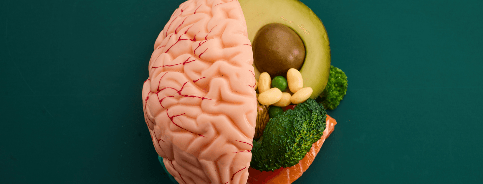
Dietary sources: Brazil nuts (highly variable but often very rich), seafood, organ meats, and eggs are among the most reliable sources in regions where soil selenium is low. Grain selenium content varies sharply by region of origin.
-
Supplementation caution: Selenium has a narrow therapeutic window. Excess selenium is toxic. Do not supplement without guidance—the goal is adequacy, not loading.
-
Children with thyroid conditions: Selenium status is worth monitoring in children with hypothyroidism or autoimmune thyroid disease, since selenoproteins protect glandular tissue and modulate immune responses relevant to autoimmunity (Triggiani et al., Endocrine, Metabolic & Immune Disorders Drug Targets, 2009).
The evidence does not yet support routine selenium supplementation for all pregnancies. What it does support is awareness: selenium is not a peripheral trace element. It is embedded in the machinery of thyroid hormone activation and brain development in ways that are only becoming clearer.
If you are pregnant, planning to become pregnant, or managing a child with neurodevelopmental concerns, a conversation with your healthcare provider about selenium—including whether testing makes sense for your situation—is a reasonable, evidence-grounded step.
References
-
Köhrle, J., & Gärtner, R. (2009). Selenium and thyroid. Best Practice & Research Clinical Endocrinology & Metabolism. https://pubmed.ncbi.nlm.nih.gov/19942156/
-
Schweizer, U., & Fabiano, M. (2022). Selenoproteins in brain development and function. Free Radical Biology & Medicine. https://pubmed.ncbi.nlm.nih.gov/35961466/
-
Ma, C., et al. (2025). Iron and selenium: At the crossroads of development and death in oligodendrocytes. Archives of Biochemistry and Biophysics. https://pubmed.ncbi.nlm.nih.gov/40517802/
-
Demircan, K., et al. (2024). Maternal selenium deficiency during pregnancy in association with autism and ADHD traits in children: The Odense Child Cohort. Free Radical Biology & Medicine. https://pubmed.ncbi.nlm.nih.gov/38704054/
-
Calcaterra, V., et al. (2025). Thyroid Health and Selenium: The Critical Role of Adequate Intake from Fetal Development to Adolescence. Nutrients. https://pubmed.ncbi.nlm.nih.gov/40732987/
-
Delange, F. (2001). Iodine deficiency as a cause of brain damage. Postgraduate Medical Journal. https://pubmed.ncbi.nlm.nih.gov/11264481/
-
Gereben, B., et al. (2008). Activation and inactivation of thyroid hormone by deiodinases: local action with general consequences. Cellular and Molecular Life Sciences. https://pubmed.ncbi.nlm.nih.gov/17989921/
-
Larsen, P. R., et al. (1995). Nutritional and hormonal regulation of thyroid hormone deiodinases. Annual Review of Nutrition. https://pubmed.ncbi.nlm.nih.gov/8527223/
-
Triggiani, V., et al. (2009). Role of iodine, selenium and other micronutrients in thyroid function and disorders. Endocrine, Metabolic & Immune Disorders Drug Targets. https://pubmed.ncbi.nlm.nih.gov/19594417/
This article is part of the Avaneuro evidence-based child development program
54 modules. 287 lessons. 140 tools. Every recommendation backed by peer-reviewed research.